


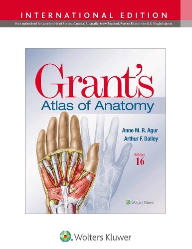
Grant's Atlas of Anatomy by Anne M R Agur Bsc Msc
Illustrations drawn from real specimens, presented in surface-to-deep dissection sequence, set Grant’s Atlas of Anatomy apart as the most accurate illustrated reference available for learning human anatomy and referencing in dissection lab. A recent edition featured re-colorization of the original Grant’s Atlas images from high-resolution scans, also adding a new level of organ luminosity and tissue transparency. The dissection illustrations are supported by descriptive text legends with clinical insights, summary tables, orientation and schematic drawings, and medical imaging.- Renowned, high-resolution, dynamically colored illustrations organized in dissection sequence enable the formation of 3D constructs for each body region and provide detailed, realistic reference during dissection.
- Tables detail muscles, vessels, and other anatomic information in an easy-to-use format ideal for review and study.
- Enhanced medical imaging includes more than 100 clinically significant MRIs, CT images, ultrasound scans, and corresponding orientation drawings to help students confidently apply the laboratory experience to clinical rotations.
- Color schematic illustrations reinforce the relationships of structures and anatomical concepts in vibrant detail.
| SKU | Unavailable |
| ISBN 13 | 9781975193447 |
| ISBN 10 | 197519344X |
| Title | Grant's Atlas of Anatomy |
| Author | Anne M R Agur Bsc Msc |
| Series | Lippincott Connect |
| Condition | Unavailable |
| Binding Type | Paperback |
| Publisher | Wolters Kluwer Health |
| Year published | 2024-02-13 |
| Number of pages | 896 |
| Cover note | Book picture is for illustrative purposes only, actual binding, cover or edition may vary. |
| Note | Unavailable |