


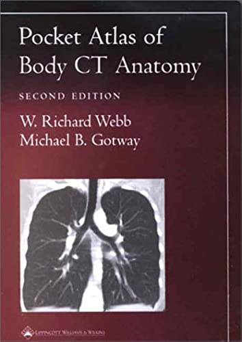
Pocket Atlas of Body CT Anatomy by W Richard Webb
Featuring 229 sharp, new images obtained with state-of-the-art technology, the Second Edition of this popular pocket atlas is a quick, handy guide to interpreting computed tomography body images. It shows readers how to recognize normal anatomic structures on CT scans...and distinguish these structures from artifacts.Chapters cover the neck and larynx, thorax, portal venous phase abdomen, pelvis, arterial phase abdomen, and reconstructions. Each page presents a high-resolution image, with anatomic landmarks clearly labeled. Directly above the image are a key to the labels and a thumbnail illustration that orients the reader to the location and plane of view. This format--sharp images, orienting thumbnails, and clear keys--enables readers to identify features with unprecedented speed and accuracy.| SKU | Unavailable |
| ISBN 13 | 9780781736633 |
| ISBN 10 | 0781736633 |
| Title | Pocket Atlas of Body CT Anatomy |
| Author | W Richard Webb |
| Series | Radiology Pocket Atlas Series |
| Condition | Unavailable |
| Binding Type | Paperback |
| Publisher | Lippincott Williams and Wilkins |
| Year published | 2002-02-12 |
| Number of pages | 144 |
| Cover note | Book picture is for illustrative purposes only, actual binding, cover or edition may vary. |
| Note | Unavailable |